Can there be anything worse than losing eyesight?
Probably nothing, but what can be equally worse is poor or damaged vision, especially if you encounter it suddenly. Of all the senses, your eyes are most valuable.
You were enjoying seeing the beautiful world and all of a sudden, everything gets distorted. This can be really dreadful. It’s almost impossible to imagine life without normal vision. Even a slight change in your eyesight can change your life drastically.
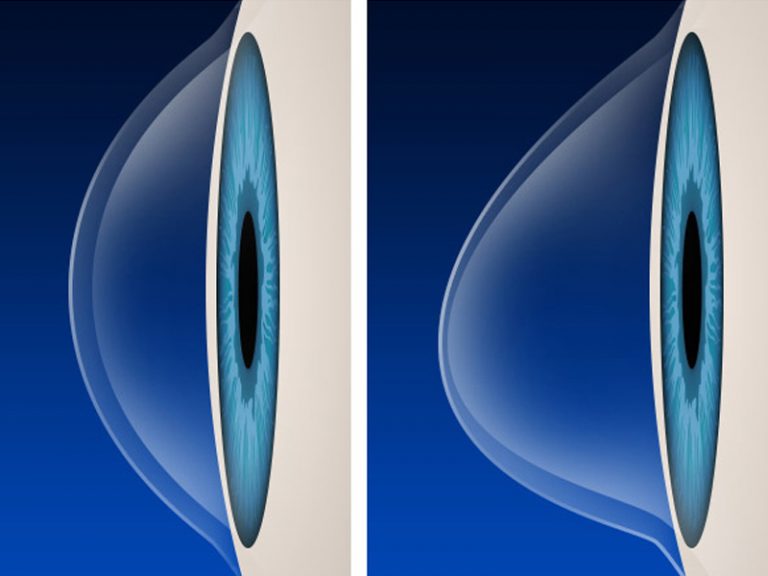
Usually it’s the cornea of your eye that can hamper the focus of the vision. According to a research, if your round cornea begins to bulge like cone shape then chances are you might be suffering from an eye disease called Keratoconus (also known as conical cornea).
This disease is not common but it’s neither rare. Out of every 2000 people, 1 is affected by Keratoconus. People in teens or early 20s are generally victimised.
Causes of Keratoconus
The real cause of Keratoconus is actually unidentified. However it is assumed that this eye disease is caused due to genetic problem or allergies.
Another cause can be imbalance of enzyme in cornea. Constantly rubbing of eyes, uncomfortable contact lenses or high exposure to UV rays can lead to Keratoconus.
Symptoms of Keratoconus
The cornea of the eyes which is normally round becomes thin, starts bulging and turns into cone-shaped. This is one of the prime signals of Keratoconus eye disease. The irregular shape of cornea often leads to:
- Slight blurring of vision
- Distorted vision
- Redness or swelling of eyes
- Lack of focus on objects
- Eyes may became more sensitive to light or glare
- Contact lenses may not fit properly
- Nearsightedness or astigmatism can also increase
Keratoconus either occurs in one eye or both the eyes. However, this defect can affect both the eyes differently.
How Keratoconus is diagnosed?
Early diagnosis of Keratoconus is really crucial to determine and maintain the status of the cornea. If Keratoconus is detected at its early stage, progression of the disease can be prevented.
Therefore as soon as you feel any change in shape of your cornea or face any of the symptoms mentioned above, get your eyes tested without further delay.
Usually the eye doctor would review patient’s medical history or ask for the family history and then proceed with the eye examination. Eye doctor or surgeon would conduct any one or more methods mentioned below to diagnose Keratoconus:
Keratoconus diagnosis
Eye refraction – Eye refraction is a general test to examine the eyes. For conducting this eye test, the doctor uses a device that measures the eyes prescription and determine the vision problem. The testing device is called phoropter or refractor, it contains different lenses. The patient is asked to look into the device to get the vision checked.
Keratometry – It is an eye test that measures the curvature of the cornea. The eye doctor uses the equipment called keratometer that determines the power of cornea on basis of the curvature measure. The basic shape of the cornea is determined and on that basis Keratoconus is checked.
Corneal mapping / topography – The computerised technique of corneal mapping is a photographic test that capture the images of the cornea and create a map that show details of the cornea shape.
In corneal topography, a map is generated which shows the surface curvature of the cornea. The thickness of the cornea can also be determined through this type of test.
Slit-lamp examination – In this test, the eye doctor uses a bright light microscope called slit lamp to check the eyes and area around the eyes. A vertical beam of light is directed into surface of the eye which helps to evaluate the shape of the cornea.
Corneal Pachymetry – It is an eye test technique in which a medical device called pachymeter is used to measure the thickness of the cornea.
Can Keratoconus be treated?
Keratoconus is not a severe disease from the initial stage itself. It take years to become worse but in some cases it can be quick too. It can turn out to be really serious if left untreated.
Treatment is mandatory for vision correction. Mild Keratoconus can be treated with the help of prescribed eyeglasses or contact lenses but if the case is crucial or if the eye disease progresses, the ophthalmologist would suggest following treatments:
Keratoconus Treatment
Corneal collagen crosslinking – It’s a treatment technique to strengthen the corneal tissue that has become thin due to Keratoconus.
The ophthalmologist uses UV lights and special eye drops to prevent the cornea from further bulging and becoming worse.
There are two types of cornea cross-linking
Epithelium-on procedure: Epi-on is actually a painless recovery process. The outer layer of the cornea is called epithelium which is removed to make way for the riboflavin (Vitamin B type) which starts functioning with UV light. Since it’s painless, you can keep going with your routine activities without any obstacle.
Epithelium-off procedure: This process is a bit painful and might require few days for recovery (around 4-5 days). During the treatment, epithelium is kept as it is and the riboflavin is penetrated due to which it requires more time.
However according to doctors, this is a safe technique as it does not cause much discomfort and also reduces the risk of infection.
Intacs – In Intacs technique, Keratoconus is treated with the help of small curved device which the ophthalmologist inserts in your cornea, surgically. Intacs are only used when the Keratoconus cannot be treated with the help of eyeglass or contact lens.
Intacs reshape the cornea and improves your vision. The surgical process takes only 10 minutes and the intacs can also be removed or exchanged.
Corneal transplant – If the condition is too serious or if the patient is unable to bare the rigidness of contact lenses, ophthalmologist strongly recommends corneal transplant.
With this technique complete or part of affected cornea is replaced by the fresh donor cornea tissue. Even after the transplant process, the patient may have to use eyeglass or contact lens for a clear vision.
Topography-guided conductive keratoplasty – When Keratoconus reaches to a stage where glasses or lenses cannot improve poor vision then topography-guided conductive keratoplasty surgery is recommended. This treatment helps to reshape the cornea without any severe complications and improve nearsightedness of patient with Keratoconus.
The energy emitted from radio waves is used to reshape the cornea. A topographic computerised map is generated of each individual patient’s eye to proceed with the treatment.
Glasses and contact lenses for Keratoconus
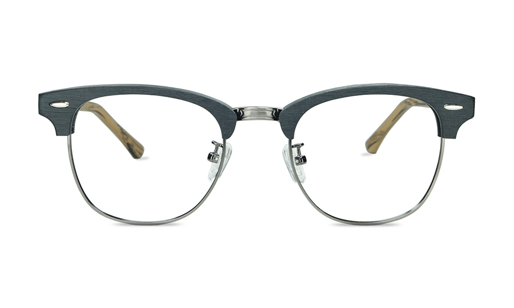
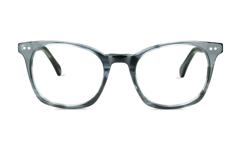
Glasses – When the Keratoconus is in its early stage, wearing proper spectacles can help to correct the shape of the cornea which is not that abnormal. Some patients with Keratoconus can notice continuous increase in astigmatism due to which they need to frequently change glasses.
Custom soft contact lenses – Along with vision clarity, patients highly demand for comfort. Usually gas permeable (GP) lenses offer discomfort. If you cannot tolerate this discomfort and demand maximum comfort for your sensitive eyes then soft contact lens is a feasible option.
The lens that fit your eyes offers clarity and comfort. Rectify corneal disorder with comfortable soft contact lens with custom parameters. The lenses can be customised as per your requirement like single vision, multifocal, toric designs etc.
Gas permeable contact lenses – If your soft contact lenses cannot control the problem of Keratoconus then doctors usually recommend Gas permeable contact lenses as it’s rigid and made up of oxygen-permeable polymers.
The lenses perfectly bend over the cornea and changes the irregular shape of the cornea into new uniform refracting surface. The hard lens perfectly rest on the top of the cornea that’s irregular and creates smooth surface which improves vision.
Hybrid contact lenses – To change the irregular shape of the cornea, hybrid contacts are specially designed that corrects the cornea.
The lenses are constructed in a combination of rigid gas permeable (RGP) surrounded by soft lens “skirt”. It offers clear vision as well as comfort of the soft lens. It provides a good fit that matches the shape of abnormal cornea caused due to Keratoconus.
Piggyback contact lenses – When Keratoconus worsens or is in mild or severe condition, soft contact lens is not a wise choice as it results in low oxygen permeability and discomfort, hence piggyback contact lens crop up as a viable choice for Keratoconus patients.
According a research where the patients with Keratoconus using these lenses, assured that Piggyback design offers maximum comfort for around 14 to 16 hours and also clear optical vision. This lens is made up of gas permeable (GP) thats fitted on the top of soft lens.
Scleral contact lenses – Scleral contact lens with large diameter is often used to correct high level of irregular astigmatism or corneal disorder caused due to astigmatism. The incredibly larger lenses rest on the sclera (the white part of the eye) and vaults present over the cornea.
The size of the scleral contacts depend upon the complexity of the corneal irregularity. Small and medium sized scleral contact lenses are available at low cost and are easy to apply whereas if the Keratoconus is advanced, larger scleral contact lenses are recommended to match the corneal curvature.
Prosthetic contact lenses – This type of lenses are used to rectify the disfigured appearance of the eye. Complex corneal disorders like Keratoconus can be corrected with Prosthetic lenses. Only if your cornea condition is critical or untreatable, Prosthetic lenses can be fitted.
Certified and trained doctors are only capable of performing this lens fitting surgery. With these lenses, the patient’s eyes will look exactly like it was before.
Stages of Keratoconus and treatment recommended
Keratoconus as said is one type of eye-disease that occurs usually between 16 to 45 years of age. However if it’s diagnosed at adolescent age, it is likely to become threatening as it could progress rapidly at least till the patient is 40 years old.
There are various stages of Keratoconus and each eye can be affected differently. Tests will be conducted to determine the severity of this cornea disorder.
Early Keratoconus
When Keratoconus is in its early stage, it does not much affect the visual condition and doesn’t tend to progress too. This eye problem can be corrected with proper prescription glasses, soft contact lenses and also with spherical or toric correction. When Keratoconus is mild the thickness of the cornea will be above 255.
Moderate Keratoconus
At this stage the corneal visibly changes and distortion increases. The low vision quality cannot be properly corrected by spectacles. For maximum comfort and clear vision, rigid gas permeable lenses are generally recommended by the eye doctors. This will improve the cornea irregularity, effectively.
However it is better to keep a pair of spectacles handy (just for emergency but you cannot be assured with 100% vision clarity), in case of loss of contacts or eye irritation. Regular checkup needs to be done to check the progression of Keratoconus and condition of the gas permeable lens or if it’s fitting well. The thickness of cornea is moderate between 475 – 524.
Advanced Keratoconus
Glasses definitely cannot cope up with the advanced stage of Keratoconus since the prescription keeps on changing. The distortion of vision increases and becomes more blurry. Scarring is observed and the cornea becomes extremely thin, hence it becomes really crucial to stabilise the condition.
Even soft, rigid gas permeable or hybrid lens cannot work effectively. For proper fitting inside the steeper curvature, miniscleral or scleral rigid gas permeable contact lenses are a good choice. Also surgical treatment is available to improve stability. The thickness of cornea majorly decreases to around 400 to 475.
Severe Keratoconus
This progressive eye disease when reaches to severe level, cornea becomes extremely thin and weakens. Corneal distortion is dramatically noticed and even corneal scarring. The sight is likely to get severely impaired at this stage. The situation progresses after 10-15 years of occurrence of Keratoconus.
Contact lenses cannot work for the severe condition as they can’t provide complete correction. Poor vision persists even after lens fitting. For severe Keratoconus stage, corneal transplant surgery is highly recommended. The cornea tremendously thins down to less than 400.
Overcome Keratoconus before it’s too late.


